What is vascular Ehlers danlos syndrome?
 Banner showing support of vEDS awareness
Vascular Ehlers Danlos Syndrome (vEDS) is a subtype of Ehlers Danlos Syndome. Ehlers Danlos Syndrome (EDS) is an umbrella term for the group of 13 different connective tissue disorders often affecting collagen function [1]. Collagen is important for skin and blood vessel strength and elasticity. In vEDS, type 3 collagen is affected which is important for the lining of blood vessel walls, skin, intestines, uterus, and lungs.
What causes veds?
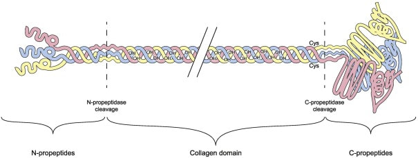
Structural domains of a type III procollagen molecule
Mutations found in the COL3A1 gene are what results in Vascular Ehlers Danlos Syndrome. COL3A1 is important for collagen type 3 synthesis by producing a
pre procollagen molecule [2].
There have been over 600 reported possible COL3A1 gene mutations, however 2/3 of them involve a glycine residue being converted into a different amino acid within the triple-helical region of the protein chain. Glycine is a critical amino acid for type 3 collagen as Glycine is present every third amino acid to help build the triple-helical weaved rope for tissue stability. Most mutations result in exon skipping and a shorter polypeptide chain, as opposed to a premature stop codon. However more research is needed to be completed to better characterize these mutations, and explore the role of collagen type 3 binding partners that also contribute to stability.
what are the signs and symptoms of veds?
|
vEDS can manifest in a variety of ways. Major symptoms include tearing or dissection of the blood vessel, uterine, and intestinal walls [3]. These events are medical emergencies and can be fatal. Many patients will experience a major event by age 30. Minor symptoms also include joint hypermobility, protruding facial features, and easy bruising.
Treatment is focused on preventative measures to reduce the risk of an emergency event. Management includes medication to control blood pressure and not engaging in high-contact sports due to tissue fragility. |
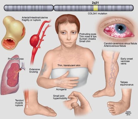
Common manifestations of vEDS
|
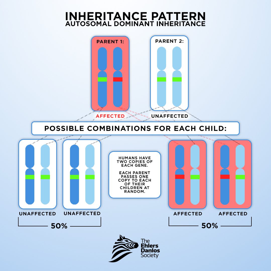
Inheritance pattern of vEDS
|
How is veds inherited?vEDS is estimated to be present in
1 in 100,000 – 200,000 people [2]. About 50% cases occur due to a de novo (non-inherited) mutation that's not caused by the genetics of the parent. The other 50% of vEDS cases are inherited in an Autosomal Dominant pattern. This means if one parent has a copy of the mutated COL3A1 gene, there is a 50% chance that each child could inherit the mutation. |
Patient Resources and advocacy groups

The Elhers danlos society
The Ehlers Danlos Society provides educational resources and advocacy opportunities for each of the combined EDS subtypes, including vEDS.
|
The VEDS Collaborative
The vEDS collaborative is a group for patients, family, researchers, and clinicians aiming to promote vEDS patient-centered research.
|

Annabelle's challenge
Annabelle's Challenge is a UK based charity that provides education and support
(including a helpline) for patients and families. |
Visitor Map
References
[1] The Ehlers-Danlos Society. (2017). What are the Ehlers-Danlos Syndromes? | The Ehlers Danlos Society. The Ehlers Danlos Society. https://www.ehlers-danlos.com/what-is-eds/
[2] Kuivaniemi, H., & Tromp, G. (2019). Type III collagen (COL3A1): Gene and protein structure, tissue distribution, and associated diseases. Gene, 707, 151–171. https://doi.org/10.1016/j.gene.2019.05.003
[3] Byers PH. Vascular Ehlers-Danlos Syndrome. 1999 Sep 2 [Updated 2019 Feb 21]. In: Adam MP, Feldman J, Mirzaa GM, et al., editors. GeneReviews® [Internet]. Seattle (WA): University of Washington, Seattle; 1993-2024. Available from: https://www.ncbi.nlm.nih.gov/books/NBK1494/
ProteinTech. (n.d.). Immunofluorescence staining of human colon tissue using a Collagen Type III Polyclonal antibody . In ProteinTech.
